SOLUTION Dermatomes and myotomes Biology Diagrams Dermatomes and myotomes cannot be overstated as they are integral parts of the human body, and understanding them can help us live healthier lives. With the proper knowledge, physical therapists, chiropractors, doctors, and surgeons can use these two structures to diagnose and treat various ailments that impair our daily routine. Furthermore

Myotomes. A myotome is a group of muscles supplied by a single spinal nerve. This is different to a motor unit, which consists of a motor neuron and the skeletal muscle fibres that it innervates. The movement-related myotome functions for the upper and lower limbs are listed below: C5: shoulder abduction and external rotation

Anatomy, Skin, Dermatomes Biology Diagrams
Dermatome And Myotome Charts - A dermatome is the location of the skin of the human anatomy that is mainly supplied by branches of a single back sensory nerve root. These spinal sensory nerves go into the nerve root at the spine, and their branches reach to the periphery of the body.

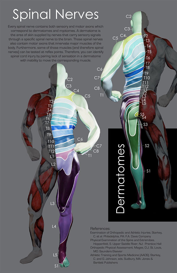
Dermatomes are areas of skin that receive sensations from sensory nerves exiting the spinal cord. Sensory nerves provide the feeling of hot, cold, pain, etc. There are 7 cervical, 12 thoracic, 5 lumbar, and 1 coccygeal nerve dermatomes. Doctors use dermatomes to help diagnose diseases and conditions. Myotomes is a group of single spinal nerves that originate from groups of muscles.
Learning Medicine, Simplified Biology Diagrams
Dermatomes are areas of skin on your body that rely on specific nerve connections on your spine. In this way, dermatomes are much like a map. The nature of that connection means that dermatomes can help a healthcare provider detect and diagnose conditions or problems affecting your spine, spinal cord or spinal nerves. Dermatomes divide the skin according to sensory nerve distribution (see Image. Dermatome Map). One of the first to map out and discuss the dermatomes is O. Foerster in his 1933 publication entitled "The Dermatomes in Man" in the journal Brain. Some consider his work the foundation of dermatomal theory.[1] In 1948, J. Keegan and F. Garrett described spinal nerve distribution in the
